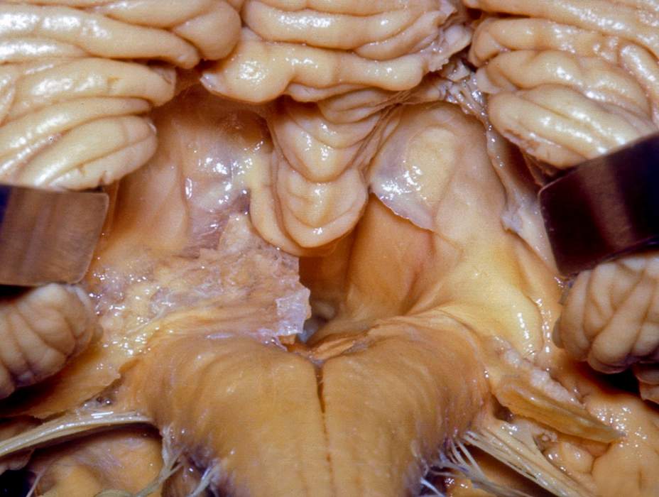
An opening in the roof of the fourth ventricle that connects to the subarachnoid space lateral apertures two openings in the side walls of the fourth ventricle that connect to the subarachnoid space.
Openings in roof of fourth ventricle.
If the opening of fourth ventricle foramen of magendie and luschka are blocked by tumor or adhesions of arachnoid mater the csf cannot enter the subarachnoid space from the ventricular cavity.
Cerebrospinal fluid escapes through this opening and lateral apertures into the subarachnoid space.
10 3 is formed by the pons and medulla fig.
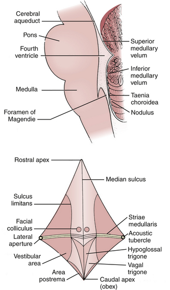
The obex is the most caudal tip of the fourth ventricle.
The upper portion of the roof is formed by the cerebellum.
Internal capsule cerebrum.
Fourth ventricle lateral walls.
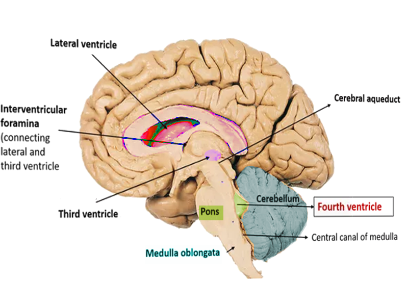
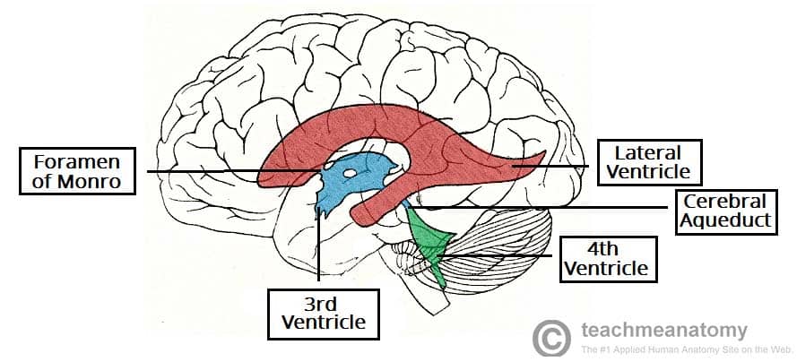
The two lateral ventricles the third ventricle and the fourth ventricle.
The roof of ventricle is diamond shaped and can be divided into superior and inferior parts.
The fourth ventricle contains choroid plexus along its roof along the tela choroidea which may protrude out the lateral foramina of luschka.
The roof of fourth ventricle is the dorsal surface of the fourth ventricle.
The roof of the fourth ventricle has presents a tent like apex at the intersection of it s superior and inferior.
The superior part of.
The fourth ventricle has a roof at its upper posterior surface and a floor at its lower anterior surface and side walls formed by the cerebellar peduncles nerve bundles joining the structure on the posterior side of the ventricle to the structures on the anterior side.
The lateral walls of the fourth ventricle are formed by the cerebellar peduncles.
The understanding of the 4th ventricle is important firstly because it s strategically set in the middle of critical structures existing in the medulla pons and cerebellum and second because its roof possesses 3 essential openings which allow the csf to escape from the ventricular system of the brain to the subarachnoid space.
This leads to excessive accumulation in the ventricular system thereby producing internal hydrocephalus.
As you look at the drawing imagine the ventricles as chambers filled with fluid.
It corresponds to the ventral surface of the cerebellum.
To the rights is a drawing of the ventricles.
As you can see the ventricles are interconnected by narrow passageways.
The sidewalls are formed by the veli and cerebellar peduncles.
It is widest at the level of the pontomedullary junction.
The lower has a median aperture foramen of magendie.
View chapter purchase book.
Roof of the fourth ventricle formed by thin laminae of white matter.
The only naturally occurring openings between the ventricles of the brain and the subarachnoid space surrounding the brain are the foramina of luschka and magendie in the fourth ventricle.
There are four in all.


:background_color(FFFFFF):format(jpeg)/images/library/13983/IxIebEKGr4ReRbXGlvvmhg_Ventriculus_quartus_01.png)


